


With severe internal diseases, poor nutrition, as well as with age, nail growth slows down, the structure undergoes changes. Only a doctor can accurately determine the cause of the disorder based on the results of tests and microscopic examinations.
But to imagine what happens to the nails on the feet or hands, you can use photos with different types of fungal diseases.
Causes of nail deformation
Forms, yeast-like fungi and dermatophyte fungi cause infectious diseases of the nail (onychomycosis) that show similar symptoms.
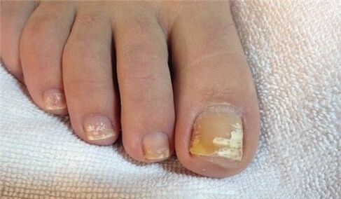
All kinds of toenail or toenail fungus deform on the nail plate, changing its transparency, shine, color, this variety can be seen in the presented photos.
Nail changes occur not only during onychomycosis, but also through lesions, chronic paronychia (inflammation of the nail fold), psoriasis, hand eczema, dermatitis. Before concluding that there is a fungal infection, you should consider all possible options.
Signs of fungal infection
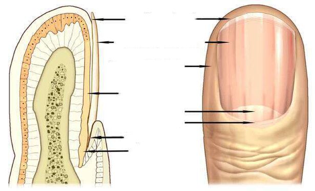
The most informative signs of a fungal infection are discoloration of the nail plate, nail separation, superficial changes - transverse, longitudinal grooves on the nail plate, point indentations, thickening, destruction of the nail.
The pink color of a healthy nail is determined by the transparency of the nail plate and the blood vessels visible through it. During onychomycosis, the nail loses transparency, the color becomes brownish, yellow, less often green, black.
Candida fungi and dermatophytes cause onycholysis - the separation of the damaged part of the nail. When infected with dermatophytes, onycholysis is observed from the distal edge of the nail, while in Candida infection, the nail falls behind the base of the nail at the base of the crescent.
A symptom of candidal fungi may be inflammation of the lateral perianal ridges - paronychia. This disease has bacterial forms caused by streptococci and staphylococci, as well as non-infectious - eczema, psoriasis, systemic vasculitis.
When the toenails are damaged by the fungus Trichophyton rubrum, the plate is affected, as you can see in the photo, the infection does not affect the roller. The plate becomes yellowish, thickens strongly, the accumulated fungal masses are well separated under it.
Nail fungus due to dermatophyte infection
In 95% of all cases of nail fungus, the disease is caused by dermatophytes Trichophyton rubrum and Trichophyton mentagrophytes.
Trichophyton rubrum infection
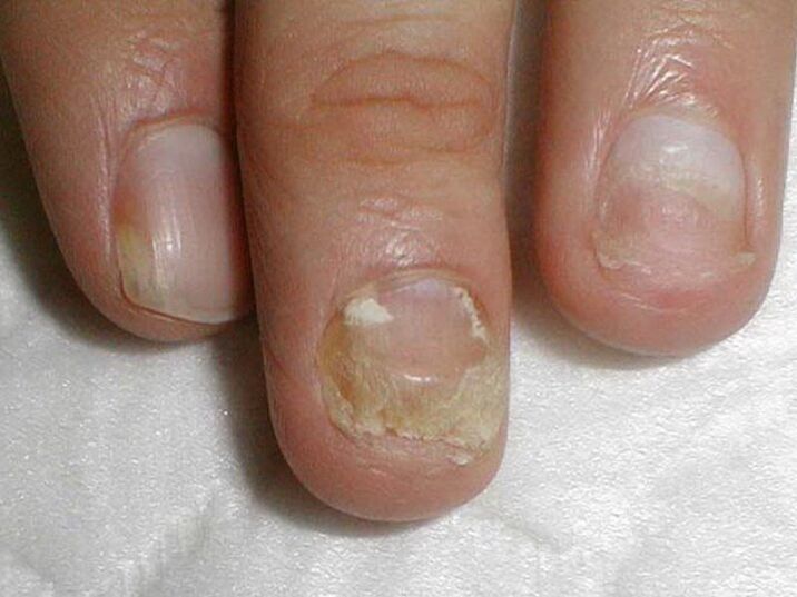
Onychomycosis begins when a fungus penetrates under the nail plate from the free edge. Fungal infection indicates yellowish varnish, uneven, disintegrated surface of the distal (distal) edge of the varnish at the site.
The distal - lateral form of the fungal infection Trichophyton rubrum dermatophyteis common. In the photo you can see that the spot caused by the introduction of the fungus is located along the fold of the lateral perianal nail.
Trichophyton rubrum The fungus usually affects the toes, causing hyperkeratosis - the accumulation of fungi between the nail plate and the nail bed, which looks like a loose yellowish mass in the photo.
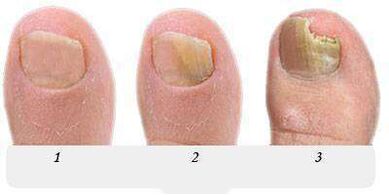
At this stage the fungus occupies a small part of the nail as shown in the photo, and with the help of local treatment it is possible to deal with onychomycosis.
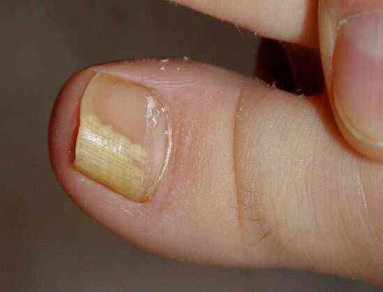
Without treatment, the spot grows, gradually affecting the entire edge of the nail and then moving to the half moon. In the photo, the nail fungus looks like a yellowish stripe pointing towards the growth zone of the nail plate.
In the fungal distal form of the nail, which is often found on the big toes, a yellowish spot of infection appears on the distal edge of the nail, in its central part, as can be seen in the photo.
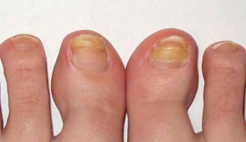
In the advanced stage of foot fungus, a few nails act as in the photo, the treatment is no longer limited to local remedies and pills. In addition to antifungal drugs, the nail is subject to technical cleaning, complete or partial removal of the nail plate.
Long-term therapy using all known antifungal agents and treatment should be carried out with foot hyperkeratosis caused by Trichophyton rubrum, as shown in the photo.
Fungal infection with complete damage to the nail spreads to the entire area of the nail plate, the nail is completely destroyed.
Infection with another dermatophyte, Trichophyton mentagrophytes, can also cause a complete fungal infection of the nail.
Infection with Trichophyton mentagrophytes
With the complete defeat of the toenail by the fungi of Trichophyton mentagrophytes, the nail plate is deformed, the photo shows that it thickens, changes structure, crumbles, yellowish spots appear on the entire surface.
Infection of the nail with this dermatophyte usually causes superficial white onychomycosis of the big toe, less often of the little finger.
This fungus is practically not found on the nails of the hands, it often causes interdigital dermatophytosis on the feet as in the photo and requires simultaneous treatment of the skin of the feet and nails.
A symptom of a fungal infection of the nail, usually on the feet, are white spots of various sizes, as in the photo, reminiscent of leukonia - a disease of the nail plate itself.
But unlike leukonia, in which white spots are caused by the formation of air bubbles in the nail layers, white spots of fungal infection are the result of the activity of Trichophyton mentagrophytes.
Rarely, superficial white onychomycosis is caused by molds; Trichophyton rubrum can cause this type of fungus in AIDS and affect the nails on both feet and hands.
Nail changes due to Candida infection
The fungus is commonly found in women, it affects the hand working on the nails, which is more often in contact with water.
Candidal onychomycosis is characterized by a proximal form of infection in which the fungus affects the nail bed at the base of the nail, then reaches the growth zone and the nail bed. It then gradually moves along the nail from the base to the edge and leads to a larger area of the nail plate.
Candida albicans is the causative agent of the disease in candidal onychomycosis. This fungus invades toenails and toenails, spreading from the half-zone of the moon at the base of the nail plate, to the free edge as shown in the photo. A sign of Candida nail infection.Candida albicans can penetrate the nail and its free edge. In this case, they are talking about the distal form of the infection, which is usually combined with candidiasis of the skin.
Treatment of Candida fungi on the nails of the hands and feet, with damage to more than half of the nail plate, as in the photo, involves not only the fight against onychomycosis, but also measures the activity of Candida in the natural reservoirs of their natural reservoirs. . .
Infection with forms
Forms are less likely to cause fungus than Candida or dermatophytes. The main symptom of a toenail infection in the form, as you can see in the photo,changes the color of the nail plate to blue, black, green.
Foot nail shape marks can be dark spots, dots on the nail plate or, as in the photo, a black longitudinal stripe.
Antifungals
Antifungals with fluconazole, ketoconazole, terbinafine, itraconazole, griseofulvin are used to treat nail fungus caused by dermatophytes, for example.
Antifungals with terbinafine are effective in treating dermatophyte infections.
Fungal agents with voriconazole are highly active against dermatophytes.
itis used andnail shapefor feet, hands andagainst candida yeast. The spectrum of action includes such forms as Aspergillium, Fusarium, Penicillium.
Itraconazole-based remedies handle molds.
Fungal-like nail diseases
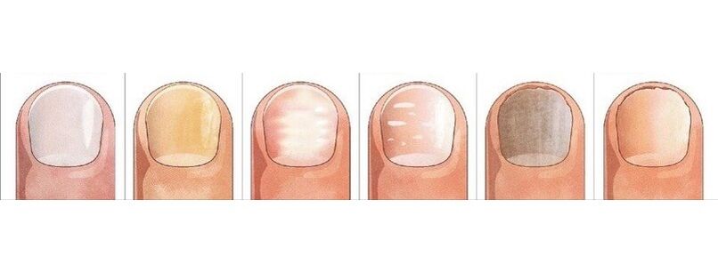
Gray tintOccasionally there is eczemaon the nail. In this case, the nail plate may move away from the nail bed, which is observed with fungus.
looks very similar to the manifestations of onychomycosispsoriasis. With this disease not onlycolor changesbut alsonail plate thickens
Spotted depressions are found on its surface, marked by the separation of the nail plate from the nail bed. But there are differences from the fungus: in psoriasis, the separated and healthy parts of the toenail are separated over time by a pink, yellowish streak.
bluish colorgets nailPseudomonas with nail infection. Frequent mechanical scratching of the nail fold causes the appearance of superficial grooves, the nail wave.
White spots of leuconiac, whose appearanceis associated with metabolic disorders, can also be mistaken for a superficial white fungus with a large area of varnish. .
Color changes, the shape of the nail causes damage. The big toes are at great risk. With nail damage, like fungus, it thickens and darkens.

The difference between an injury and a fungus is that changes in the lesion are noted only on the injured finger, the nails on the other fingers remain intact, not infected from the diseased finger as in onychomycosis.
The result of trauma can be a partial separation of the nail from the nail bed, creating a cavity that under unfavorable conditions quickly colonizes with fungi.
It is possible to separate the nail plate from the nail bed under the influence of light (photonicolysis), iron deficiency anemia, hormonal diseases. Split nail, loss occurs with lichen erythematosus, bullous dermatoses, nail trauma.
But finally make sure the conclusion is correct and start treatment, only a dermatologist - a dermatologist or a mycologist - a doctor treating fungal diseases can help.